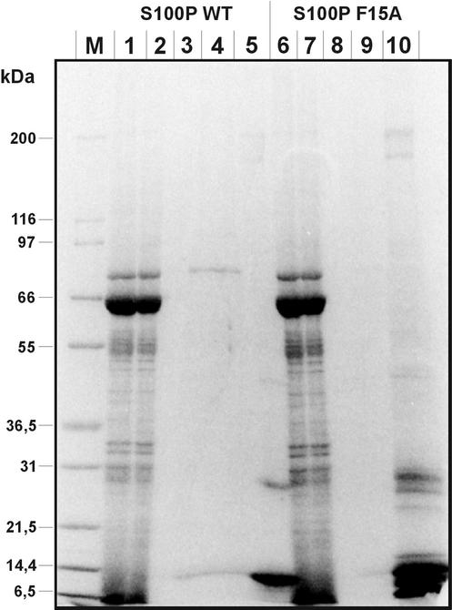
Figure 1.
Affinity chromatography of placental proteins on immobilized WT S100P (lanes 1–5) and F15A S100P (lanes 6–10). Placental protein extract (lanes 1 and 6) was loaded onto each column in a Ca2+-containing buffer. The flow through was collected (lanes 2 and 7), and columns were then washed in the presence of Ca2+ (lanes 3 and 8). Subsequently Ca2+-dependently bound proteins were eluted with a buffer containing EGTA (lanes 4 and 9). Finally the columns were treated with an imidazole-containing buffer to strip off all bound proteins (lanes 5 and 10). M, molecular weight markers. Proteins present in the different fractions were subjected to SDS-PAGE and then stained with Coomassie. Note the presence of an 80-kDa polypeptide in the EGTA eluate of the WT S100P column (lane 4), which is not found in the F15A S100P eluate (lane 9).

