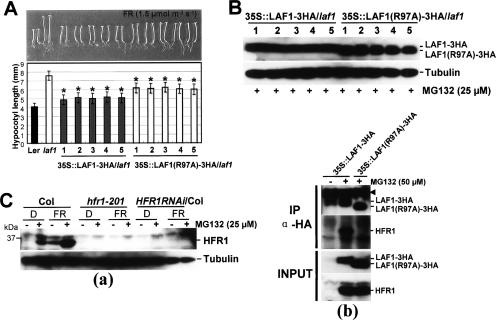
Figure 5.
Phenotypes of transgenic Arabidopsis seedlings expressing LAF1 or LAF1(R97A) in laf1. (A) Complementation of the laf1 mutant by LAF1 or LAF1(R97A)-3HA overexpression under FR light. Five independent transgenic lines were analyzed for each construct. LAF1 #1 (n = 100), #2 (n = 100), #3 (n = 100), #4 (n = 80), and #5 (n = 80), and LAF1(R97A) #1 (n = 100), #2 (n = 100), #3 (n = 100), #4 (n = 100), and #5 (n = 100) seedlings were grown for 4 d under FR light (1.5 μmol m−2 sec−1) on media without sucrose. Data are presented as the average hypocotyl length ± standard deviation (SD). (*) LAF1-overexpressing transgenic seedlings are significantly shorter than LAF1(R97A) overexpression seedlings (P < 0.01, Student’s t-test). (B) Western blot analysis of transgenic Arabidopsis seedlings overexpressing LAF1-3HA (lanes 1–5) or LAF1(R97A)-3HA (lanes 1–5) under FR irradiation with MG132. After treatment with MG132 (25 μM), seedlings were incubated for 12 h under continuous FR light (1.5 μmol m−2 sec−1). Tubulin levels were used as loading controls. Note that LAF1(R97A)-3HA has a faster mobility than LAF1-3HA. As these samples were analyzed on the same blot, their signal strengths can be directly compared. (C) Endogenous HFR1 levels in wild type and laf1. (Panel a) Western blot analysis of wild type (Col), hfr1-201, HFR1RNAi/Col using anti-HFR1 antibody. Seedlings grown in darkness for 4 d were transferred to FR light (1.5 μmol m−2 sec−1) or darkness with or without MG132 (25 μM) for 12 h. Tubulin levels were used as loading controls. (Panel b) Coimmunoprecipitation of endogenous HFR1 with LAF1 protein. Protein extracts of transgenic Arabidopsis seedlings [35S∷LAF1-3HA or 35S∷LAF1(R97A)-3HA] treated with MG132 (50 μM) under FR light (1.5 μmol m−2 sec−1) for 12 h were immunoprecipitated with anti-HA. Four-day-old 35S∷LAF1-3HA seedlings grown in darkness were used as a control. Input proteins and the immunoprecipitates were separated on 10% SDS–polyacrylamide gels, blotted onto membranes, and detected with anti-HA and anti-HFR1 antibodies. Input refers to the starting protein amount in extracts used for immunoprecipitation reactions. The arrowhead indicates the cross-reaction with the heavy chain of the protein A-conjugated antibody.

