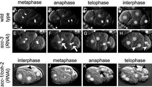
Figure 1.
Depletion of each cohesin component homolog causes defect in chromosome segregation. Time-lapse images of early embryos of wild-type (A–D), scc-3 (RNAi) (E–H), and scc-1/coh-2 (RNAi) (I–L) embryos. These images can be seen also in video movies (WT.mov, scc-3.mov, and coh-2.mov, respectively). Fluorescence of GFP-tagged histone H2B and the DIC images are overlaid. The arrows in F and K indicate anaphase chromosomes with abnormal morphology. The arrowheads in H, I, and L indicate small extranuclei generated by abnormal chromosome segregation. Bar, 5 μm.

