Figure 4.
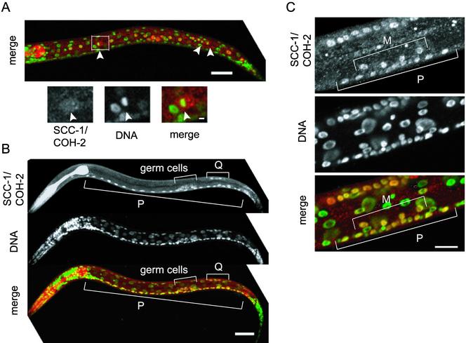
SCC-1/COH-2 is expressed in dividing cells throughout the development. (A) An L1 larva stained with anti-SCC-1/COH-2 antibodies (SCC-1/COH-2, red) and Sytox Green (DNA, green). The SCC-1/COH-2 signal was intense in the 14 hypodermal V lineage cells, which divide synchronously. The magnified view shows a cell at metaphase in which the SCC-1/COH-2 signal is dispersed in the cytoplasm. Arrowheads indicate condensed chromosomes. Bar, 20 μm. (B) A slightly older L1 larva, stained as described above. In addition to the dividing P lineage and Q lineage cells, germ cells were stained specifically with anti-SCC-1/COH-2 antibodies. Bar, 20 μm. (C) The ventral region of an L3 larva. The SCC-1/COH-2 signal could be detected in 4 M lineage and 10 P lineage cells, which divide concurrently. Bar, 10 μm.

