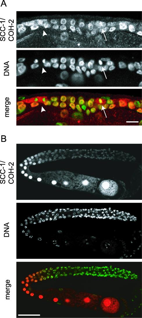
Figure 5.
SCC-1/COH-2 localizes to nuclei in germ cells. Hermaphrodite germ cells were stained with anti-SCC-1/COH-2 antibodies (SCC-1/COH-2, red) and Sytox Green (DNA, green). (A) Mitotic germ cells in an L3 larva. As in somatic cells, SCC-1/COH-2 was dispersed in the cytoplasm of germ cells at prometaphase (arrows), and was not detected on condensed anaphase chromosomes (arrowheads). Bar, 10 μm. (B) In a gonad of an adult hermaphrodite, SCC-1/COH-2 was weakly detected on the chromosomes of mitotic germ cells. The SCC-1/COH-2 signal became very intense in maturing oocytes and was spread evenly in the nuclei. Bar, 50 μm.

