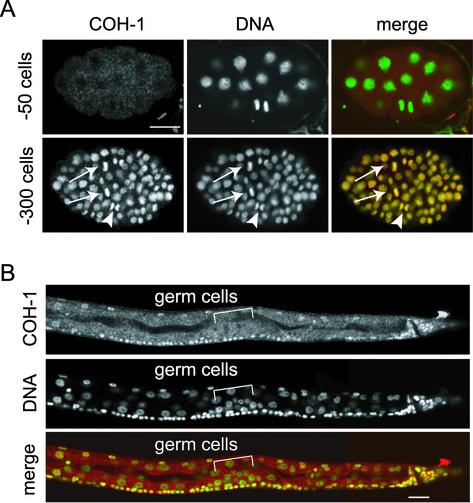
Figure 6.
Localization of COH-1 in embryos and larvae. (A) Wild-type embryos stained with anti-COH-1 antibody (COH-1, red) and Sytox green (DNA, green). COH-1 was not detectable in an early embryo (∼50 cells). In an embryo of a later stage (∼300 cells), COH-1 associated chromosomes in every cell, regardless of the phase of the cell cycle, including metaphase (arrows) and anaphase (arrowheads). Bar, 10 μm. (B) The posterior half of an L2 larva, stained as described above. COH-1 localized to chromosomes of virtually all somatic cells, but was missing from the germ cell nuclei. Bar, 10 μm.

