
Fig. 1.
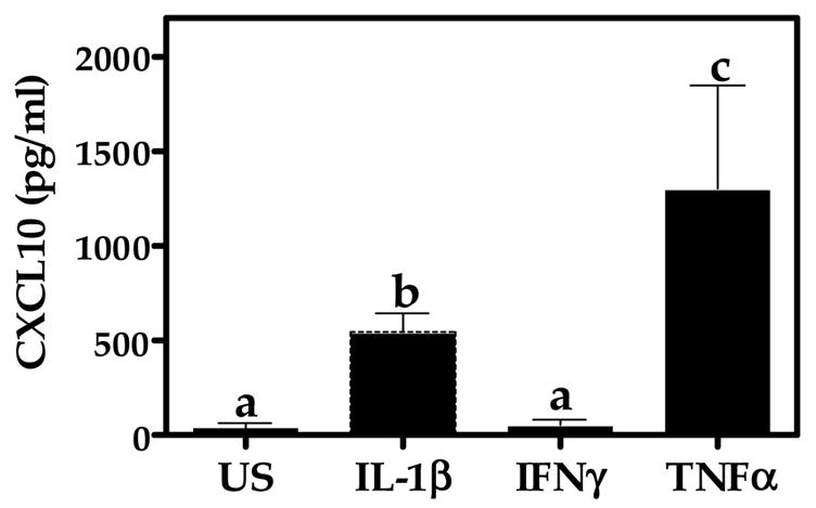
Cytokine-induced CXCL10 protein expression in A172 cells. Astroglial cells were unstimulated (US) or exposed for 24 h to human recombinant TNFα, IL-1β, or IFN-γ (5 ng/ml). A standard dual-antibody solid phase immunoassay (ELISA) was used for quantitation of secreted CXCL10 in cell culture supernatants. Data represent mean + S.E.M. of duplicate measures from 3 independent experiments. Data points with any common letters above them are not statistically different from each other as determined by one-way analysis of variance (ANOVA) with Neuman-Kuels’ multiple comparisons.