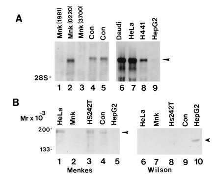
Figure 1.

(A) RNA blot analysis of Menkes mRNA in cell lines from Menkes patients (lanes 1–3), control primary fibroblasts (lanes 4 and 5), and human cell lines (lanes 6–9). Ten micrograms of total RNA was isolated, electrophoresed, transferred to nylon, and hybridized with a human Menkes [32P]cRNA probe. (B) Immunoblot of cell lysates with Menkes (lanes 1–5) or Wilson (lanes 6–10) antibody. Protein was loaded at 75 μg per lane, and after SDS/10% PAGE proteins were transferred to nitrocellulose and analyzed by ECL (Amersham).
