
Figure 5.
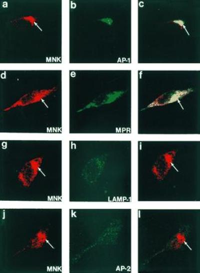
Confocal immunofluorescence localization of the Menkes ATPase in HeLa cells. Cells were processed for indirect immunofluorescence and analyzed after incubation with antibodies to the Menkes ATPase (a, d, g, and j) and γ-adaptin (AP-1) (b), the cation-independent mannose-6-phosphate receptor (MPR) (e), lamp-1 (h), and AP-2 (k). The combined confocal image from each double-labeling is shown in c, f, i, and l. (×400.)