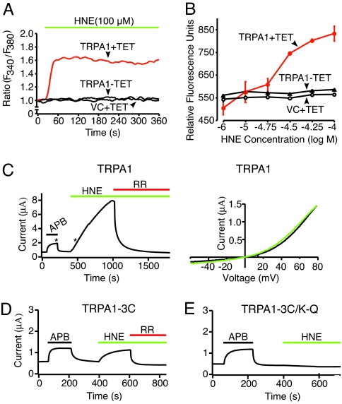
Fig. 1.
HNE activates the cloned TRPA1 channel. (A) HEK293 cells expressing rat TRPA1 after tetracycline induction (TRPA1+TET; red trace) were challenged with HNE (100 μM), and responses was assessed by calcium imaging. Uninduced (TRPA1-TET; black trace) or vector-transfected control cells (VC+TET; black trace) showed no significant response. (B) Dose–response analysis of HNE-evoked calcium responses in tetracycline-induced (TRPA1+TET; red), uninduced (TRPA1-TET) or vector-transfected control (VC+TET) HEK293 cells. Analysis was performed by using a multiwell fluorescence plate reader. Each well contained 30,000 cells; n = 4 wells per agonist concentration. (C) Representative voltage-clamp recording (+70 mV holding potential; Left) from Xenopus oocytes expressing human TRPA1 channel, showing robust activation by HNE and block by ruthenium red (RR). Asterisks indicate times at which voltage ramps were acquired to generate current–voltage relationships (Right) for APB (black) and HNE (green). (D and E) Oocytes expressing TRPA1–3C or TRPA1–3C/K-Q mutant channels were challenged with 2-aminoethyl diphenylborinate (APB; 200 μM; black), HNE (200 μM; green), or ruthenium red (RR; 10 μM; red), as indicated.

