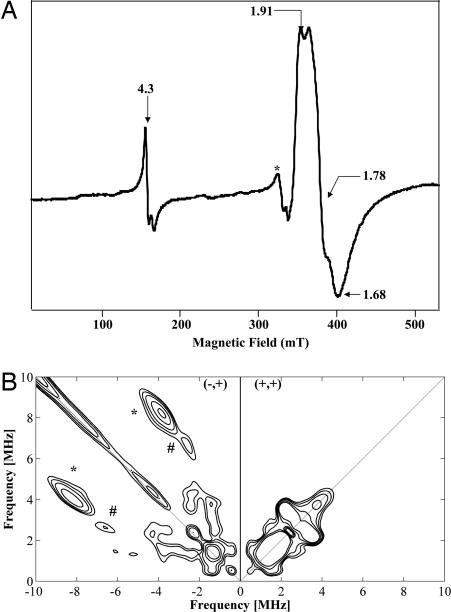
Fig. 3.
Characterization of MiaE2H protein by EPR spectroscopy. (A) X-band EPR spectrum of the MiaE2H protein (1.9 mM) in 100 mM Tris·HCl (pH 8) containing 30 mM NaCl and 5% glycerol. Experimental conditions: temperature 4 K, microwave power 1 mW, modulation amplitude 10 mT. The weak signal indicated by a star at g ≈ 2.00 is contaminating Cu2+. (B) Low-frequency region of the X-band HYSCORE spectrum of the mixed-valent state [FeII–FeIII] of MiaE2H protein (1.9 mM) in 100 mM Tris·HCl (pH 8) containing 30 mM NaCl and 5% glycerol. Experimental conditions: magnetic field 3,900 G (g = 1.778), frequency 9.7 GHz, and temperature 4 K.

