Fig. 4.
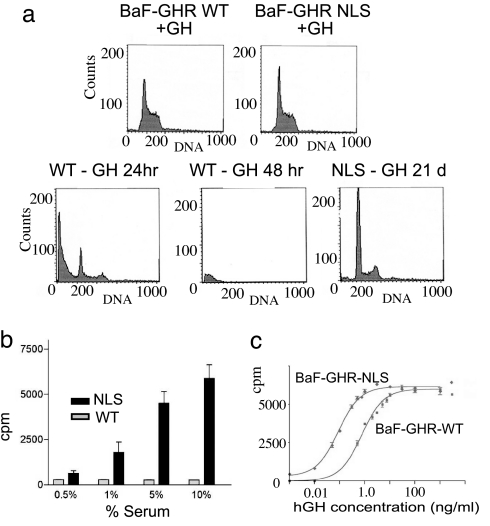
Effect of increased GHR nuclear targeting in a cell model. In a subconfluent culture with GH support, both BaF-GHR WT and NLS lines undergo mitosis, with 60% in G1 phase and 40% in S/M phase (a Upper). However, upon the withdrawal of cytokine support (in the form of GH or IL-3), only the BaF-GHR NLS lines proliferate (a Lower; only 21-day sort shown for NLS line, exemplifying earlier timepoints). (b) BaF-GHR NLS lines continue to proliferate in the absence of GH and IL-3, with the support of fetal bovine serum. (c) NLS-GHR lines show a dramatic increase in the sensitivity to GH as measured by [3H]thymidine incorporation assay (20-fold decrease in ED50, i.e., 10 pM for NLS, compared with 200 pM for WT in 1.0% serum). Results were confirmed in three separate population studies and clonal lines from three separate transfections.

