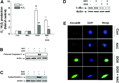
Figure 6. Effects of NAC on DOX-triggered cell death signalling.
Cells were treated with 4 μM DOX in the absence or presence of 5 mM NAC for different time periods (24 h for ROS, 48 h for cytochrome c, 12 h for IκBα and 16 h for NF-κB p65 respectively) and then the amount of intracellular ROS, caspase-3, cytochrome c, IκBα and NF-κB p65 were determined as described in the Materials and methods section. (A) NAC inhibited DOX-mediate ROS generation. Values are means±S.E.M. for three independent experiments (##P<0.01 compared with control; **P<0.01 compared with DOX alone). (B) NAC inhibited DOX-mediated caspase-3 cleavage. (C) NAC inhibited DOX-mediated enhancement of cytosolic cytochrome c levels. (D) NAC failed to inhibit DOX-mediated IκBα degradation. (E) NAC was unable to inhibit DOX-mediated NF-κB p65 translocation. The images are representative of three independent experiments yielding similar results. Green, NF-κB p65; dark blue, nucleus; white, NF-κB p65 in the nucleus.

