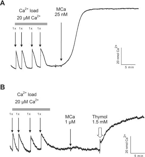
Figure 5. MCa fails to induce Ca2+ release from cardiac HSR vesicles.
Representative traces of extravesicular Ca2+ concentration variations measured using Antipyrylazo III, a Ca2+ indicator. Skeletal HSR vesicles (A) and cardiac HSR vesicles (B) were actively loaded with Ca2+ by sequential addition of 20 μM CaCl2 (final concentration) in the cuvette. After each Ca2+ addition, the absorbance was monitored until the initial baseline value was reached. MCa-induced Ca2+ release was then tested on skeletal and cardiac HSR vesicles by addition of 25 nM MCa to skeletal HSR (A) and 1 μM MCa to cardiac HSR (B). Integrity of the cardiac vesicles and functionality of the RyR2 channel were assessed by injection of 1.5 mM thymol, 10 min after the application of MCa.

