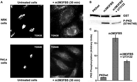
Figure 3. PI-PLC activator m-3M3FBS induces Golgi fragmentation and PKD1 activation.
(A) NRK and HeLa cells were incubated respectively with 25 and 50 μM m-3M3FBS for 30 min. Then, the integrity of TGN structure was analysed by immunofluorescence. (B) Lysed extracts from GST–PKD1-transfected HeLa cells were analysed by Western blot to measure PKD1 activation. In some experiments, cells were incubated with U73122 before and during m-3M3FBS treatment (third lane from left). (C) Densitometry of the experiment described in (B). Values are means (±S.D., vertical bars) for three separate experiments.

