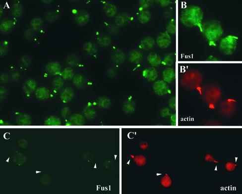
Figure 8.
Indirect immunofluorescence localization of Fus1 to the fertilization tubule of activated mt+ gametes. (A) Anti-Fus1 indirect immunofluorescence of wild-type mt+ gametes. (B and B′) Corresponding micrographs of wild-type mt+ gametes dual-labeled with the Fus1 polyclonal antibody (B, green) and the filamentous actin-specific fluorochrome Alexa 546-phalloidin (B′, red). (C and C′) Corresponding micrographs of fus1-1 mt+ gametes dual-labeled with the Fus1 polyclonal antibody (C) and the actin-specific fluorochrome Alexa 546-phalloidin (C′). The arrowheads point to the position of fertilization tubules.

