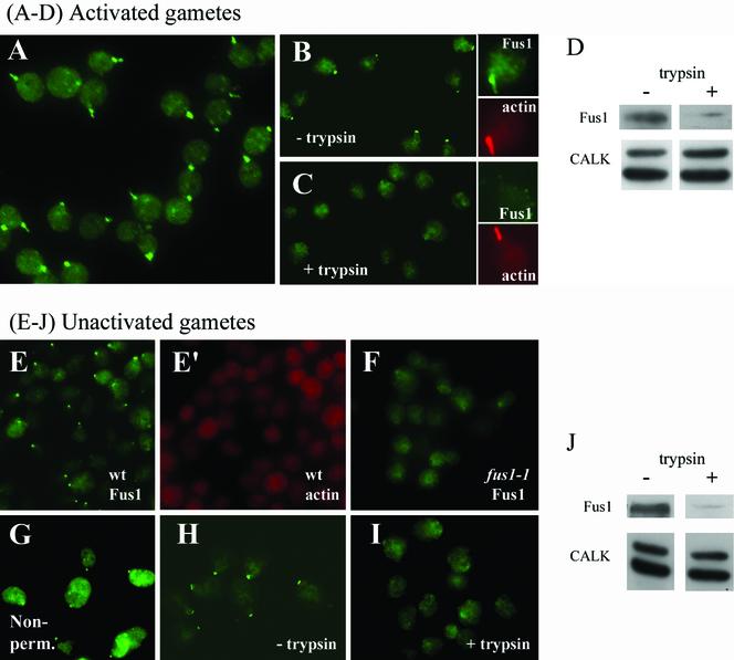
Figure 9.
Fus1 is on the external surface of fertilization tubules in activated wild-type mt+ gametes and in a discrete patch on the external surface of unactivated wild-type mt+ gametes. (A) Fluorescence micrograph of activated gametes not permeabilized before immunolocalization with the Fus1 antibody. (B and C) Activated wild-type mt+ gametes incubated with 0% (B) or 0.5% trypsin (C). The insets for B and C each show control (B) and trypsin-treated (C) samples dual labeled with anti-Fus1 antibody (green) and Alexa 546-phalloidin (red). (D) Activated wild-type mt+ gametes incubated with 0.05% trypsin were analyzed for Fus1 by immunoblotting (top). Identical samples also were immunoblotted for CALK, an intracellular protein (bottom). In each lane, 2 × 10⁷ cells were loaded. (E and E′) Corresponding micrographs of unactivated wild-type mt+ gametes dual labeled with the anti-Fus1 antibody (E, green) and the actin-specific fluorochrome, Alexa 546-phalloidin (E′, red). (F) Unactivated fus1 mt+ gametes incubated with the anti-Fus1 antibody. (G) Indirect immunofluorescence image of unactivated nonpermeabilized wild-type mt+ gametes stained with the anti-Fus1 antibody. (H and I) Anti-Fus1 indirect immunofluorescence images of control (H) and 0.5% trypsin-treated (I) unactivated wild-type mt+ gametes. (J) Anti-Fus1 immunoblot of unactivated wild-type mt+ gametes treated with 0.05% trypsin (top). The lower panel shows an anti-CALK immunoblot of identical samples.

