Figure 5.
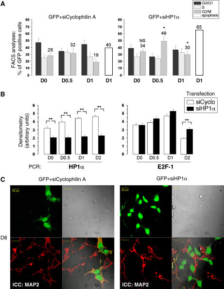
HP1α knockdown impairs proper neuronal differentiation and activates e2f-1 expression. P19 cells were co-transfected with pGFP and siRNA directed against cyclophilinA as control or HP1α, 72 h before differentiation induction. (A) Time course analyses of the transfected cell population by FACS. One-way ANOVA test followed by Bonferroni means comparison test was performed. The G2/M values for each time points of siHP1α/GFP-transfected cells were compared to the corresponding times of siCyclophilin A/GFP-transfected cells. * indicates statistical difference compared to control cells with P<0.05 (B). Expressions of e2f1 and hp1α genes in P19 cells transfected with control siRNA against cyclophilin A and HP1α were measured by semiquantitative RT–PCR. Bands were quantified and results are presented as arbitrary units of densitometry. One-way ANOVA test followed by Bonferroni means comparison test was performed. The values for each time points of siHP1α-transfected cells were compared to the corresponding times of siCyclophilin A-transfected cells. ** indicates statistical significance with P<0.01. (C) Immunocytochemistry at day 8 (D8) after differentiation with MAP2 antibody (red) and visualization of GFP-expressing cells (green). The GFP-transfected cells (that previously showed a transient knockdown for HP1α at D1, see Supplementary Figure S3D,E) are not MAP2-positive.

