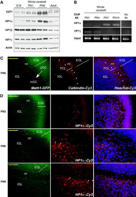
Figure 6.
Expression and binding of HP1 proteins in vivo. (A) Cerebelli were dissected at different developmental stages of the mice: embryonic stage 18 (E18), PN1, PN6 and adult (PN50). Western blot analysis was performed with antibodies against E2F1, HP1α, β, γ and actin as indicated. (B) Cerebelli were dissected from mice at PN1, PN4, PN7 and PN10 and ChIP experiments were performed on fresh tissue with antibodies against HP1α or HP1γ with primers specific to the e2f1 gene promoter. Control PCR experiments were performed with total DNA (input) and DNA isolated in the absence of primary antibody, no Ab. A typical experiment is shown. (C) Cerebellar sections from Math1-GFP transgenic mice at PN9 were immunostained for calbindin (middle panel). Most intense GFP was found in the EGL reflecting the normal expression pattern of Math1 in pGC (left panel). An overlay of Hoechst staining and Cy3 immunolabeling is presented on the right panel. The arrow indicates pGC migration through ml to IGL where mGC are localized. Asterisks show single Purkinje cells in PCL. Scale bar 50 μm. (D) Cy3-coupled HP1α immnunostaining was performed on cerebellar sections from Math1-GFP transgenic mice at different PN as noted. Indications are the same as in (C). Note the increased population of HP1α-positive mGC in the IGL at PN6.

