
Fig. 4.
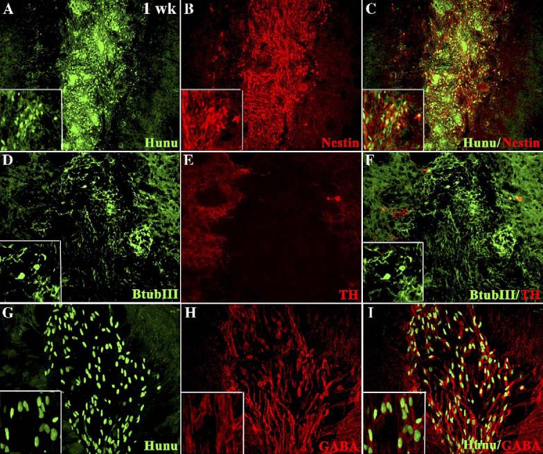
Characterization of NP-hMSC spheres 1 week after transplantation into the striatum of 6-OHDA-treated rats. Immunocytochemical localization of hunu (A), nestin (B), and images merged in panel C, β-III tub (D), TH (E), and images merged in panel F, hunu (G, GABA (H) and images merged (I)). High-power (40×) insets are included in panels A–D, F, G–I.