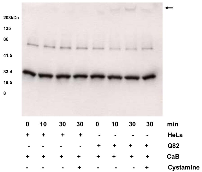
Figure 3.

Calbindin-D 28k (CaB) Western blot showing TG2 catalyzed time-dependent crosslinking of CaB with ataxin-1. HeLa cell lysates expressing GFP and GFP-Q82 ataxin-1 were used. No insoluble CaB aggregates are seen in lanes containing GFP expressing HeLa cell lysates. In contrast, lysates of Q82 expressing cells showed insoluble deposits containing CaB on top of the stacking gel (arrow). The aggregate formation was inhibited by 10 mM cystamine.
