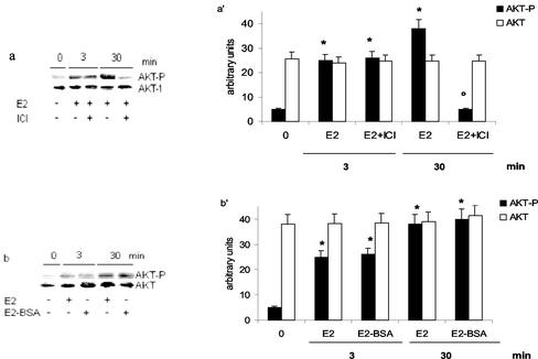
Figure 3.
Involvement of estrogen receptor in E2-induced AKT phosphorylation in HepG2 cells. Western blot analyses of AKT phosphorylation were performed as described in MATERIALS AND METHODS in HepG2 cells stimulated with vehicle (-) or with 17β-estradiol (E2) (10 nM) for 3 and 30 min with or without 15 min of pretreatment with the pure antiestrogen ICI 182,780 (ICI) (1 μM) (a) or on E2–BSA-stimulated cells (10 nM) for 3 and 30 min (b). The same filters were reprobed with anti-AKT antibody. a′ and b′, densitometric analysis (means ± SD) of three independent experiments. *p < 0.001 compared with respective control values (0), determined using Student's t test. °p < 0.001 compared with respective estradiol values (E2), determined using Student's t test.

