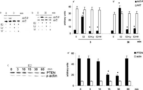
Figure 4.
Effect of E2 on AKT upstream signaling molecules in HepG2 cells. Involvement of PI3K in E2-induced AKT phosphorylation was studied by Western blot analysis as described in MATERIALS AND METHODS on HepG2 cells treated with the PI3K inhibitors Ly 294002 (Ly) and wortmannin (W) (10 μM each) and stimulated for 3 or 30 min with vehicle (0) or estradiol (E2) (10 nM) (a and b). Time course of PTEN levels (c) was analyzed by Western blot on control (0) and on E2-treated (10 nM) HepG2 cells at different times. The same filters were reprobed with anti-AKT or anti–β-actin antibodies. a′, b′ and c′, densitometric analysis (means ± SD) of three independent experiments. *p < 0.001 compared with respective control values (0), determined using Student's t test. °p < 0.001 compared with respective E2 values (E2), determined using Student's t test.

