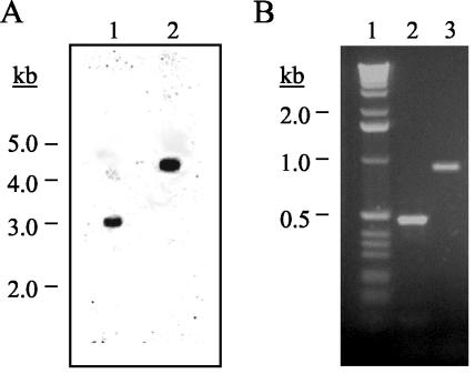
FIG. 2.
Analysis of the PAC1 disruption strain. (A) Southern analysis. Genomic DNA (2 μg) from the wild-type strain (lane 1) and strain PAC2A (PAC1 disruption strain) (lane 2) was digested with HindIII, separated by electrophoresis in a 0.7% agarose gel, transferred to a nylon membrane, and probed with a 32P-labeled DNA fragment of PAC1. The molecular size standards (kilobases) are indicated. (B) PCR Analysis. PAC2A genomic DNA was amplified with primer pairs pacD5:h3p (lane 2) and pacD3:h5p (lane 3). The molecular size standards (kilobases) are shown in lane 1. The DNA was separated by electrophoresis in a 1% agarose gel. The gel was stained with ethidium bromide and photographed under UV illumination.

