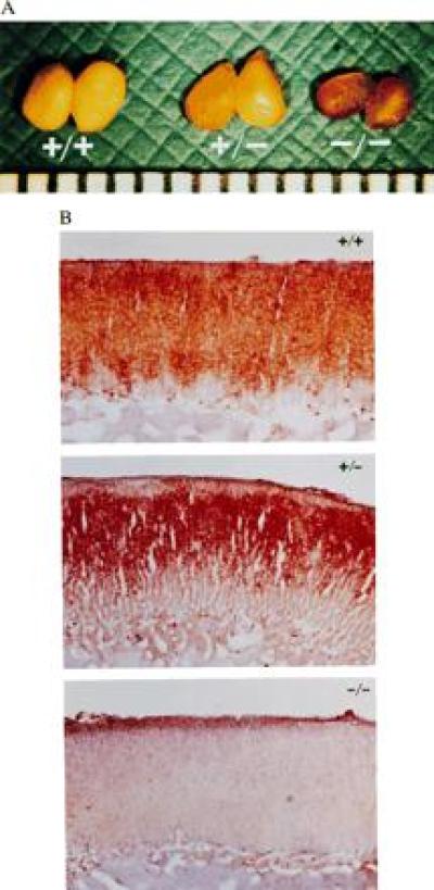
Figure 2.

Adrenal lipid depletion in Acact−/− mice. (A) Gross appearance of adrenal glands from male Acact+/+, Acact+/−, and Acact−/− mice. The scale at the bottom of the photograph is in millimeters. (B) Oil red O-stained sections showing absence of neutral lipids in adrenal cortex of male Acact−/− mice. Identical findings were observed in female mice. Staining in the heterozygous mice was consistently confined to a smaller area of the zona fasciculata.
