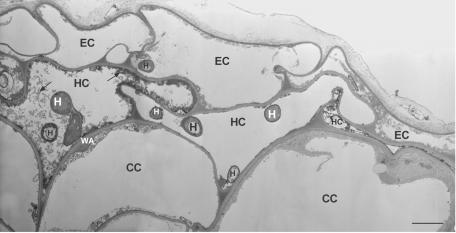
FIG. 3.
TEM images of ultrathin cross sections showing colonization of flax root tissues at 4 days postinoculation with the pathogenic strain F. oxysporum Foln3. Colonization of epidermal and hypodermal cells and the defense reactions (osmiophilic material [arrow], wall appositions, and collapsed cells) are similar but less intense than those observed in root tissues colonized by the nonpathogenic strain Fo47 (see Fig. 2). Scale bar, 2.5 μm. Abbreviations: CC, cortical cell; EC, epidermal cell; H, hypha; HC, hypodermal cell; WA, wall apposition.

