
Figure 1.
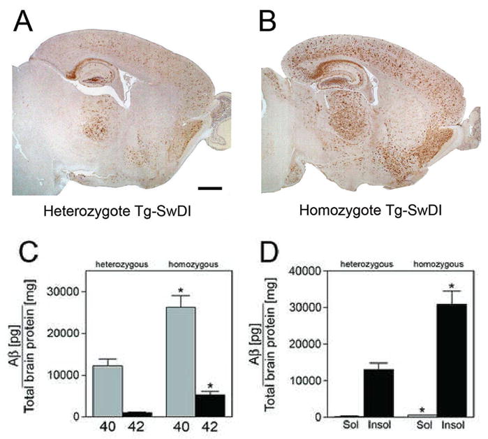
Homozygous Tg-SwDI mice accumulate markedly more Aβ than heterozygous animals. Aβ immunostaining in twelve months old heterozygous (A) homozygous (B) Tg-SwDI reveals more extensive deposition in the latter. Scale bar = 1 mm. (C) Total Aβ 40 and Aβ 42 loads in homozygous Tg-SwDI mice is more than double compared to heterozygous animals. *P < 0.001. (D) The levels of soluble Aβ and insoluble Aβ are similarly increased in homozygous Tg-SwDI mice compared with heterozygous animals. Data presented are the mean ± S.D. (n = 10 per group). *P < 0.0005.