
Figure 5.
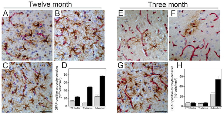
Elevated numbers of reactive astrocytes in the subiculum of young Tg-SwDI mice. Microvascular-associated reactive astrocytes revealed by GFAP-positive immunostaining (brown) and collagen type IV for identification of microvessels (red) in the fronto-temporal cortex (A,E), thalamus (B,F), and subiculum (C,G) from twelve months old and three months old Tg-SwDI mice, respectively. Scale bars = 50 μm. Quantitative stereological measurement of reactive astrocyte densities in different brain regions of twelve months old (D) and three months old (H) wild-type (gray bars) or Tg-SwDI mice (black bars). Data shown are the mean ± S.D. (n = 6 mice per age group).