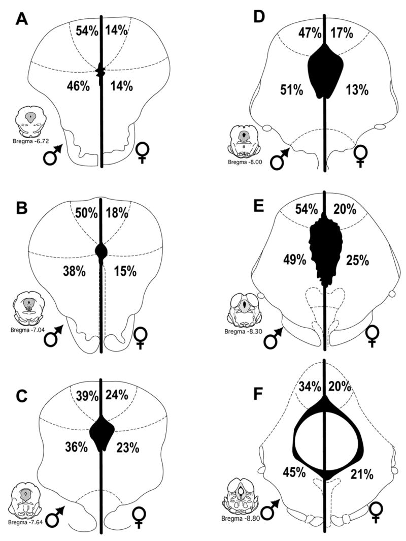
Figure 5.

Percentage of morphine-induced Fos+ PAG neurons that were retrogradely labeled from the RVM (Fos+FG) in male (left) and female (right) rats in the dorsomedial and lateral/ventrolateral regions of the PAG at six representative levels. The proportion of Fos positive neurons projecting to the RVM was greater in male than female rats across all PAG regions.
