Figure 1.
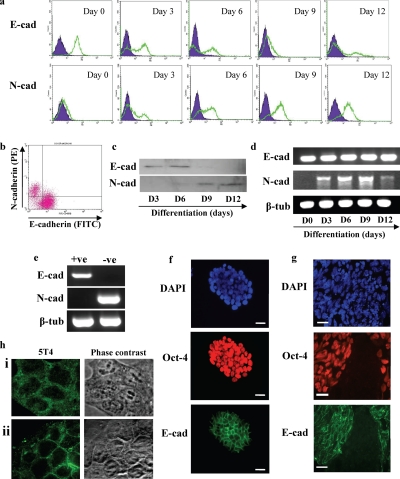
Mouse ES cell spontaneous differentiation is associated with loss of cell surface E-cadherin protein and gain of cell surface N-cadherin protein. MESC20 ES cells were maintained in an undifferentiated state by culture in ES cell medium containing LIF and differentiated in synthetic serum in the absence of LIF in monolayer culture. (a) Cell surface E-cadherin (E-cad) and N-cadherin (N-cad) proteins were assessed in undifferentiated MESC20 ES cells (day 0) and differentiated cells for 3, 6, 9, and 12 d by fluorescent flow cytometry in a Becton-Dickinson FACScaliber. E- or N-cadherin, open population; isotype control antibodies, closed population. (b) Fluorescent flow cytometry dual staining for E- and N-cadherin on MESC20 ES cells differentiated for 3 d as described above. (c) Western blot was performed to assess total cellular E- (E-cad) or N-cadherin (N-cad) proteins in MESC20 ES cells differentiated for 3, 6, 9, and 12 d as described above. (d) RT-PCR analysis of E- (E-cad) and N-cadherin (N-cad) and β-tubulin (β-tub; control) transcript expression was assessed in undifferentiated MESC20 ES cells (day 0) and in cells differentiated for 3, 6, 9, and 12 d as described above. (e) MESC20 ES cells were differentiated for 3 d as described above and E-cadherin–positive (+ve) and E-cadherin–negative (−ve) cells isolated by FACS. RT-PCR was performed on the samples to assess E- (E-cad) and N-cadherin (N-cad) and β-tubulin (β-tub) transcript expression. (f) Immunofluorescence microscopy analysis of total cells (DAPI) and E-cadherin (E-cad) and OCT-4 protein expression in undifferentiated MESC20 ES cells. Bar, 10 μm. (g) MESC20 ES cells were differentiated for 4 d and assessed for total cells (DAPI) and E-cadherin (E-cad) and OCT-4 protein expression using immunofluorescent microscopy. Bar, 10 μm. (h) ES cells were cultured in FCS+LIF (i) and FCS−LIF (ii) for 2 d and 5T4 antigen expression assessed using fluorescent microscopy and phase-contrast microscopy.

