Figure 6.
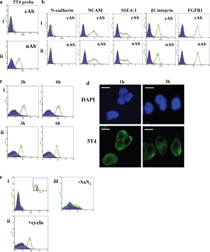
Loss of E-cadherin–mediated cell–cell contacts in wild-type ES cells results in transcriptional- and translational-independent localization of the 5T4 antigen at the cell surface. Undifferentiated D3 or MESC20 ES cells were cultured on gelatin-treated plates in FCS+LIF. (a) D3 ES cells were cultured in the presence of (i) control antibody (cAb) or (ii) E-cadherin–neutralizing antibody DECMA-1 (nAb) and 5T4 antigen expression assessed by fluorescent flow cytometry analysis. 5T4 antigen (open population); isotype control (closed population). (b) Undifferentiated D3 cells were cultured in FCS+LIF and the presence of (i) cAb or (ii) nAb and assessed for expression of the cell surface proteins N-cadherin, NCAM, SSEA-1, β1-integrin, and FGFR1. Note that no significant differences were observed for these antigens in the two antibody treatments. Similar results were obtained with MESC20 ES cells and E-cadherin null ES cells (data not shown). (c) (i) Undifferentiated MESC20 ES cells were cultured in the presence of cAb (filled population) or nAb (open population) for 3 and 6 h, and cell surface 5T4 antigen was assessed by fluorescent flow cytometry. (ii) Cell surface 5T4 antigen expression was determined in MESC20 ES cells after removal of cAb or nAb from the cells for 3 and 6 h using fluorescent flow cytometry. (d) Undifferentiated MESC20 ES cells were cultured in the presence of nAb for 1 and 3 h and 5T4 antigen (5T4) assessed by fluorescent microscopy. DAPI shows all cell nuclei within the field of view. Bar, 5 μm. (e) (i) ES cells expressing GFP under control of the 5T4 promoter were cultured in the presence of cAb (closed population) or nAb (open population) for 3 h, and GFP expression was assessed using fluorescent flow cytometry. Inset shows GFP expression in these cells after removal of LIF for 3 d. (ii) MESC20 ES cells were cultured in the presence of cAb (closed population) or nAb (open population) and 10 μg/ml cyclohexamide to inhibit total protein synthesis, and cell surface 5T4 antigen was assessed using fluorescent flow cytometry after 3 h. (iii) MESC20 ES cells were cultured in the presence of cAb (closed population) or nAb (open population) and 10 μM sodium azide to inhibit energy (ATP)-dependent processes and cell surface 5T4 antigen assessed using fluorescent flow cytometry after 3 h. Similar results were also obtained with D3 ES cells (data not shown).

