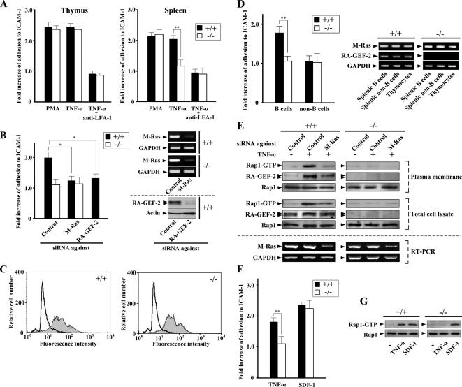
Figure 6.
Impaired LFA-1–mediated adhesion to ICAM-1 in RA-GEF-2−/− cells from the spleen, but not the thymus. (A) Comparison of adhesion to ICAM-1 between cells from wild-type and RA-GEF-2−/− mice. Thymocytes and splenocytes isolated from wild-type and RA-GEF-2−/− mice were left untreated or treated with PMA and TNF-α. After treatment, cells were examined for adhesion to ICAM-1. Anti-mouse LFA-1 antibody was added 30 min before TNF-α treatment. Fold increase of adhesion to ICAM-1 of cells treated with PMA or TNF-α compared with that of untreated cells was shown. The percentages of adhesion to ICAM-1 of untreated cells are as follows: wild-type thymocytes, 7.6%; RA-GEF-2−/− thymocytes, 7.3%; wild-type splenocytes, 9.7%; and RA-GEF-2−/− splenocytes, 5.9%. Each bar represents the average and SE of six independent experiments performed in triplicate. ■, wild-type mice; □, RA-GEF-2−/− mice; **p < 0.01. (B) Inhibition of TNF-α–induced adhesion to ICAM-1 by down-regulation of M-Ras and RA-GEF-2 in splenocytes. Splenocytes isolated from wild-type and RA-GEF-2−/− mice were transfected with siRNAs against control (Control), M-Ras, and RA-GEF-2 and left untreated or treated with TNF-α. After treatment, cells were examined for adhesion to ICAM-1. Fold increase of adhesion to ICAM-1 of cells treated with TNF-α compared with that of untreated cells is shown. The percentages of adhesion to ICAM-1 of untreated cells are follows: wild-type splenocytes, 9.2%; RA-GEF-2−/− splenocytes, 6.3%. Each bar represents the average and SE of three independent experiments performed in triplicate. ■, wild-type mice; □, RA-GEF-2−/− mice; *p < 0.05. Reduction of the expression of M-Ras and RA-GEF-2 in wild-type (+/+) and RA-GEF-2−/− (−/−) splenocytes by specific siRNAs was confirmed by RT-PCR and immunoblotting, respectively. (C) Equal expression of LFA-1 in wild-type and RA-GEF-2−/− mice. LFA-1 on the cell surface was stained with fluorescein isothiocyanate-conjugated anti-mouse LFA-1 (M17/4) antibody and analyzed by FACScaliber. White- and gray-colored histograms represent the isotype control, and cells stained with anti-LFA-1 antibody, respectively. (D) TNF-α–induced adhesion of splenic B- and non-B-cells. Splenic B- and non-B-cells isolated from wild-type and RA-GEF-2−/− mice were left untreated or treated with TNF-α. After treatment, cells were examined for adhesion to ICAM-1. Fold increase of adhesion to ICAM-1 of TNF-α–treated cells compared with that of untreated cells was shown. The percentages of adhesion to ICAM-1 of untreated cells are as follows: wild-type B-cells, 5.8%; RA-GEF-2−/− B-cells, 4.6%; wild-type non-B-cells, 11.7%; RA-GEF-2−/− non-B-cells, 11.3%. Each bar represents the average and SE of four independent experiments performed in triplicate. ■, wild-type mice; □, RA-GEF-2−/− mice; **p < 0.01. The expression level of M-Ras and RA-GEF-2 in splenic B-cells, splenic non-B-cells, and thymocytes from wild-type (+/+) and RA-GEF-2−/− (−/−) mice was confirmed by RT-PCR. (E) The activation of Rap1 and RA-GEF-2 recruitment in the plasma membrane fraction in response to TNF-α. Splenocytes isolated from wild-type and RA-GEF-2−/− mice were transfected with siRNAs against control (Control) and M-Ras, and extracts from the plasma membrane–enriched fraction were prepared after TNF-α treatment. The GTP-bound form of endogenous Rap1 in the plasma membrane fraction and the total cell lysate was precipitated from the extracts by the use of GST-RalGDS-RID and detected by immunoblotting using anti-Rap1 antibody. The amount of endogenous RA-GEF-2 and Rap1 in the plasma membrane–enriched fraction was monitored by immunoblotting using anti-RA-GEF-2 and anti-Rap1 antibodies, respectively. The amount of endogenous RA-GEF-2 and Rap1 in total cell lysates was also monitored by anti-RA-GEF-2 and anti-Rap1 antibodies, respectively. Inhibition of M-Ras expression by the siRNA in total cell lysates was confirmed by RT-PCR using primers specific for M-Ras. (F) Adhesion to ICAM-1 of splenocytes in response to TNF-α and SDF-1. Splenocytes isolated from wild-type and RA-GEF-2−/− mice were treated with TNF-α (10 ng/ml) and SDF-1 (100 ng/ml) for 30 and 5 min, respectively, and examined for adhesion to ICAM-1. Fold increase of adhesion to ICAM-1 of cells treated with TNF-α or SDF-1 compared with that of untreated cells was shown. The percentages of adhesion to ICAM-1 of untreated cells are as follows: wild-type splenocytes, 7.7%; RA-GEF-2−/− splenocytes, 5.3%. Each bar represents the average and SE of three independent experiments performed in triplicate. ■, wild-type mice; □, RA-GEF-2−/− mice; ** p < 0.01. (G) The activation of Rap1 in splenocytes treated with TNF-α and SDF-1. Splenocytes isolated from wild-type and RA-GEF-2−/− mice were treated with TNF-α and SDF-1 as in F. After treatment, the GTP-bound form of endogenous Rap1 was precipitated from total cell lysates by the use of GST-RalGDS-RID and detected by immunoblotting using anti-Rap1 antibody. The amount of endogenous Rap1 in total cell lysates was monitored by anti-Rap1 antibody.

