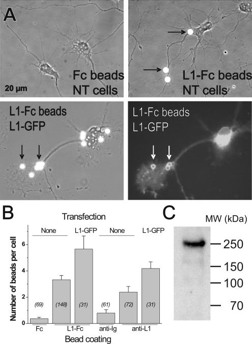
Figure 4.
Specific binding of L1–Fc and anti-L1–coated microspheres to neurons. (A) Untransfected neurons (NT) or neurons transfected for L1–GFP were incubated for 0.5 h with latex microspheres coated with human Fc, L1–Fc, or anti-L1 antibodies, and then they were rinsed and fixed. Note the accumulation of L1–GFP fluorescence around L1–Fc- or anti-L1–coated microspheres (white arrows). (B) The number of beads bound per cell in each condition is expressed as mean ± SEM, with the number of cells examined in italics. (C) The purified L1–Fc protein was run on a polyacrylamide gel and immunoblotted with antibodies against L1, showing migration at the expected molecular weight.

