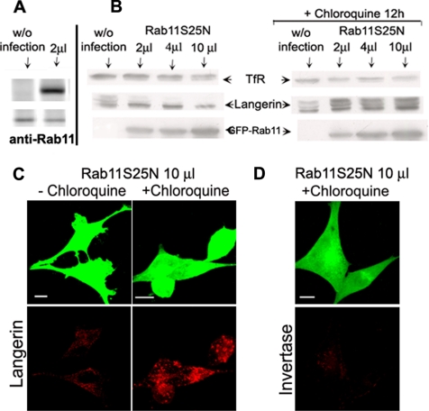
Figure 3.
Effects of Rab11AS25N overexpression on Langerin levels in M10-22E cells. Immunoblotting was used to compare the levels of TfR, Langerin, and GFP–Rab11A in uninfected cells and cells infected with increasing amounts (2, 4, or 10 μl) of Rab11AS25N adenoviruses, in the presence (B, right) or absence (B, left) of 50 μM chloroquine, as described in Materials and Methods. Proteins were separated by SDS-PAGE in 12% acrylamide gels and blotted onto membranes, with equivalent amounts of protein being loaded in each lane. The positions of TfR, Langerin, and GFP–Rab11A are indicated by arrows. Blots are representative of three independent experiments with individual blots being probed for all three proteins. As a control for endogenous Rab11 versus GFP–Rab11AS25N expression, both proteins were probed with anti-Rab11 antibodies in uninfected cells (without infection) and cells infected with 2 μl of GFP–Rab11AS25N adenoviruses (A). M10-22E cells infected with 10 μl of Rab11AS25N were incubated with (C, right, and D) or without (C, left) 50 μM chloroquine, as described in Materials and Methods. The cells were then immunolabeled with the anti-Langerin mAb DCGM4 (C) or incubated with Cy3-invertase for 1 h at 37°C before fixation (D).

