Figure 4.
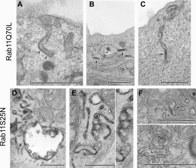
Effects of Rab11A recombinant adenoviruses on the distribution and presence of Birbeck granules in M10-22E cells. M10-22E cells were transduced with recombinant adenoviruses encoding GFP–Rab11AQ70L (20 μl [?] of stock solution) (A–C) or GFP–Rab11AS25N (2 μl of stock solution) (D–F), in the presence of 10 mg/ml HRP and/or a gold-labeled anti-Langerin mAb during the last 30 min of infection. An open-ended “BG-like structure” appended to the cell surface and coated over its entire length (arrowheads) is shown in A. Cytosolic BGs (arrows) are essentially located at the cell periphery, close to the plasma membrane. They are either isolated (B) or continuous with vacuolar endosomal structures (star) (B) and tubular membrane structures (C). As shown in D and E, tubular and vacuolar HRP+ networks occur in M10-22E cells expressing Rab11AS25N, with variable proportions of each type of network between cells. Thus, the vacuolar component is readily seen in D, whereas the tubular component predominates in E. These networks are linked to the early endosomal pathway, as shown by the occasional presence of gold-labeled anti-Langerin mAbs (a gold-labeled area in E is circled and enlarged on the right-hand side). In contrast, BGs are absent from these tubular networks. Only very occasionally (F), small tubular structures resembling BGs (arrow) with a very short, thin, central striation and a surrounding coat (arrowheads) can be seen, either apparently isolated in the cytosol or connected to the tubular network. Bars, 0.5 μm.

