Figure 3.
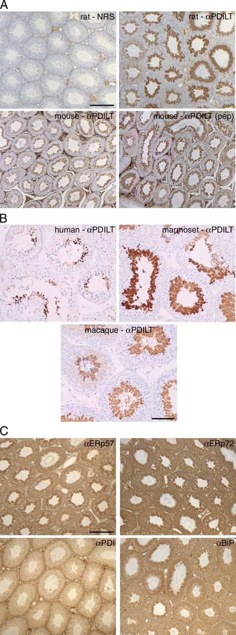
Expression of PDILT is germ cell specific. (A) Sections from rat testis counterstained with hematoxylin (blue) were immunostained with normal rabbit serum (NRS) or an anti-PDILT serum raised against recombinant PDILT at a dilution of 1:8000 (top panels). The PDILT staining pattern (brown) is similar for mouse testis (bottom left) or when using a serum raised against an internal PDILT peptide at 1:1000 (bottom right). Scale bar, 200 μm. (B) Testis sections from human, macaque, and marmoset were immunostained for PDILT. Although a single population of PDILT-positive germ cells occupied the full circumference of the tubule in macaque, the staining on human and marmoset was more heterogenous consistent with the existence of tubules containing more than one stage of spermatogenesis in a single cross section. Scale bar, 100 μm. (C) Rat testis sections were immunostained for ERp57, ERp72, PDI, and BiP (brown). Expression of these proteins was observed throughout the seminiferous epithelium. Scale bar, 200 μm.

