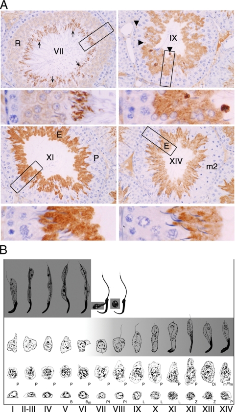
Figure 4.
Germ cell expression of PDILT from stage VII. (A) Rat testis sections immunostained with anti-PDILT from Figure 3A were imaged at higher magnification. Four representative sections from stages VII, IX, XI, and XIV of the rat spermatogenic cycle are shown. A higher detailed section, indicated by a box, is shown below each stage. Round spermatids (R), elongate spermatids (E), and immunonegative pachytene spermatocytes (P) and germ cells undergoing meiotic division (m2) are indicated. At stage VII mature elongate spermatids ready for release are indicated by arrows. At stage IX immunopositive staining was particularly intense in small areas (arrowheads) that are likely to be the residual cytoplasmic remnants from spermatozoa released at stage VII. (B) A staging diagram showing the germ cell associations characteristic of the spermatogenic cycle (Russell et al., 1990) summarizes the expression levels of PDILT (gray shading) in the adult rat.

