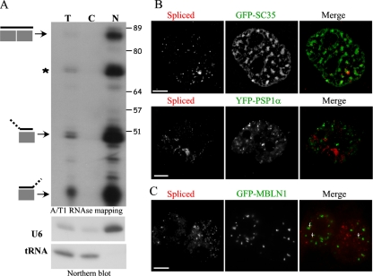
Figure 5.
Bsr RNA, a novel nucleus-restricted poly(A) RNA. (A) Subcellular fractionation of REFs. Top, cytoplasmic versus nuclear fractions represent 81 versus 19% of the total, respectively. The same absolute amount of RNA (10 μg) was loaded in order to visualize enrichment in any fraction (cytoplasmic or nuclear) that would contain Bsr RNAs, compared with the input. Bottom, the quality of the fractions was checked by Northern blot using tRNA and U6 snRNA as cytoplasmic and nuclear markers, respectively. Note that the bands indicated by an asterisk (*) and those corresponding to hemi-protected Bsr exons can be due to alternative RNA splicing and/or sequence polymorphisms. Size (nt) is indicated. (B) Distribution of spliced Bsr RNA species relative to nuclear speckles (top) and paraspeckles (bottom) that are visualized by GFP-SC35 and YFP-PSP1α staining, respectively. To highlight speckle domains, pictures have been processed using a high-pass filter. Bar, 5 μm. (C) Distribution of spliced Bsr species relative to CUG-repeat foci made by mutant DMPK transcripts. REFs were transiently transfected with a mutant DMPK mini-gene containing 960 CUG repeats in its 3′ untranslated region and a GFP-MBNL1–expressing plasmid. Expanded CUG repeat-containing transcripts form RNA foci are visualized by GFP-MBNL1 signals (green). Small white arrows indicate the few Bsr foci (detected by a Cy3-labeled probe, red) that overlap with those made by CUG foci.

