Figure 1.
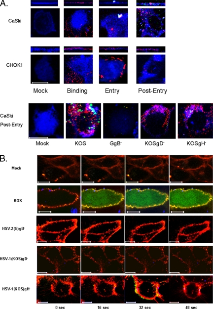
HSV enters CaSki, but not CHO-K1, cells, and it elicits an increase in both membrane and global intracellular Ca2+. (A) CaSki or CHO-K1 cells were exposed to DiD-envelope–labeled HSV-1(KV26GFP) virus (moi = 10 pfu/cell) in a synchronized assay, and then they were examined by confocal microscopy. Plasma membranes stained with EZ-Link are blue, DiD-stained viral envelopes are red, and GFP-tagged viral capsid proteins are green. Bottom, CaSki cells were mock infected or exposed to DiD-labeled wild-type or deletion viruses (moi = 10 pfu/cell for wild-type and an equivalent number of viral particles for the deletion viruses) in a synchronized assay, fixed, permeabilized, and stained for the viral capsid protein VP5 (green) 1 h after entry. Areas of colocalization between cell membrane and envelope look turquoise. Images are representative of 100 cells. Bar, 10 μm. (B) CaSki cells were loaded with Calcium Green and mock infected or infected with each of the DiD-labeled viruses. Live images acquired every 2 s beginning immediately after the temperature reached 37°C. For these images, the plasma membrane is red, viral envelopes are blue, and Ca2+ is green; colocalization of Ca2+ and plasma membrane is yellow.

