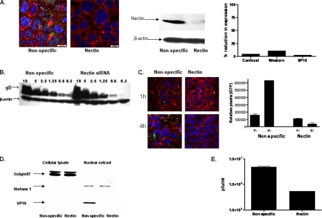
Figure 3.
Blockade of nectin-1 by siRNA prevents viral entry after binding. (A) Cells were transfected with nonspecific or nectin-1–specific siRNA and 48 h after transfection, they were stained with a cocktail of anti-nectin-1 Abs (green) and examined by confocal microscopy (plasma membranes, red; nucleus, blue) (left). Alternatively, cell lysates for Western blotting were prepared 48 h after transfection, and then they were incubated with anti-nectin mAbs, stripped, and reprobed with mAb to β-actin. Representative blots from three independent experiments are shown (middle). Blots and confocal images were quantified as described for Figure 2. (B) Transfected CaSki cells were exposed to serial dilutions of HSV-2(G) at the moi indicated for 5 h at 4°C. The cell-bound viral particles were detected by analyzing Western blots of cell lysates for gD; blots were also probed with a mAb to β-actin to control for cell loading. The blot is representative of three independent experiments. (C) Transfected cells were infected with K26GFP virus (moi = 5) and 1 and 4 h after infection, the cells were fixed, stained, and viewed by confocal microscopy (red; plasma membrane, blue, nuclei; green, viral capsids). Results are representative of three independent experiments. The relative amount of intracellular GFP was compared by collecting data from ∼100 cells from different fields using NIH Image J software. Results are expressed as the pixel intensity per cell in relative units. (D) Transfected CaSki cells were exposed to HSV-2(G) (moi = 1) in a synchronized infection assay, and cell lysates and nuclear extracts were prepared 1 h after infection and analyzed for presence of viral tegument protein VP16 by Western blotting. Blots were also probed for the cytosolic protein, golgin 97, and the nuclear protein, histone H1 Results shown are representative of three independent experiments. Blots were scanned, and nuclear VP16 detected in nectin-siRNA–transfected cells as a percentage of VP16 detected in nonspecific siRNA-transfected cells was compared (A, right). (E) The transfected cells were exposed to serial dilutions of HSV-2(G). Plaques were counted 48 h pi, and viral titer was determined. Results are means ± SD of three independent experiments conducted in duplicate.

