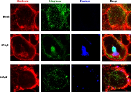
Figure 5.
Interactions with integrinαv requires viral gH. Transfected CaSki cells were mock infected or infected with purified DiD-envelope labeled gD− or gH− virus (viral particle numbers equivalent to moi = 10 pfu/cell) in a synchronized assay and fixed and stained for integrinαv 15 min after the temperature reached 37°C. Areas of colocalization between integrinαv (green) and viral envelopes (blue) are turquoise, and areas of colocalization between the plasma membrane (red) and viral envelope (blue) are purple. Results are representative of those obtained with ∼100 cells in five independent experiments.

