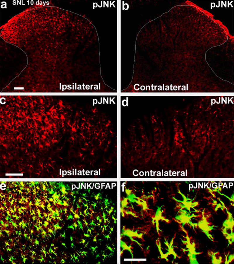
Fig. 2. SNL induces persistent JNK activation in spinal astroglia.

(a,b) Immunohistochemistry reveals an increase in pJNK in the ipsilateral spinal dorsal horn (L5) 10 days after SNL. White lines indicate the border of the dorsal horn. Scale, 50 μm. (c,d) High-magnification images of (a) and (b), respectively, showing pJNK staining in the medial superficial dorsal horn. Scale, 50 μm. (e) Double immunofluorescence shows that pJNK (red) colocalizes with the astroglial marker GFAP (green) in the medial superficial dorsal horn. Two single-stained images are merged. c, d, e have the same magnification. (f) High-magnification image of (e) demonstrates colocalization of pJNK and GFAP. Note that some fine processes of astrocytes are labeled by pJNK but not by GFAP antibody. Scale bar, 25 μm. Reproduced, with permission, from (Zhuang et al., 2006a).
