
Figure 1.
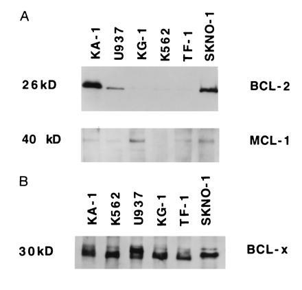
Expression of BCL-2, MCL-1 and BCL-x proteins in myeloid leukemia cell lines. (A) Whole cell lysates from Kasumi, U937, KG-1, K562, TF-1 and SKNO-1 cells were separated on a 12% SDS/PAGE. The gel was stained with Coomassie blue after transfer to demonstrate equal loading of proteins (not shown). The levels of BCL-2 were detected by Western blot analysis, using antibodies specific for BCL-2 proteins. The membrane was stripped of bound antibody and reprobed with an antibody specific for MCL-1. (B) The cell lysates used in Fig. 1A were tested for the presence of the BCL-x protein by Western blot analysis, using antibodies that specifically recognize BCL-x protein.