
Figure 3.
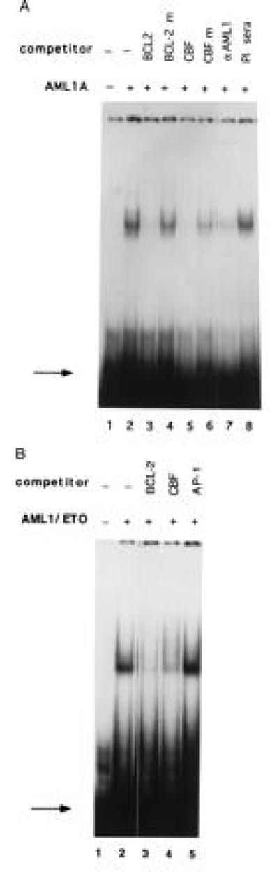
AML1A and AML1/ETO bind to the BCL-2 promoter. (A) A 32P-labeled oligonucleotide containing the AML1 binding site from the BCL-2 promoter was run alone (lane 1), or incubated with 5 μl of bacterially expressed AML1A protein (lane 2). The reaction mixture was preincubated with a 100-fold molar excess of unlabeled wild-type oligonucleotide (lane 3) or an oligonucleotide with a mutated AML1-binding site (lane 4), an oligonucleotide containing the AML1 site from the IL-3 promoter (lane 5) or the IL-3 promoter oligonucleotide with a mutated AML1 site (lane 6). In lane 7, the AML1A protein was preincubated with 2 μl of AML1 antiserum, and in lane 8 with 2 μl of preimmune serum. The binding of AML1A to the BCL-2 promoter was analyzed by EMSA. The position of the free probe is indicated by the arrow. (B) The 32P-labeled 40-mer oligonucleotide (shown in Fig. 2) was incubated with 5 μl of bacterially expressed AML1/ETO alone (lane 2), or with a 100-fold molar excess of unlabeled BCL-2 oligonucleotide (lane 3), a 100-fold excess of unlabeled oligonucleotide containing the AML1 binding site from the IL-3 promoter (lane 4) or a 100-fold excess of unlabeled oligonucleotide containing a consensus AP-1 site (lane 5). No protein was added in lane 1. The position of the free probe is indicated by the arrow.