
Figure 1. ErbB-4 is expressed in GABAergic interneurons.
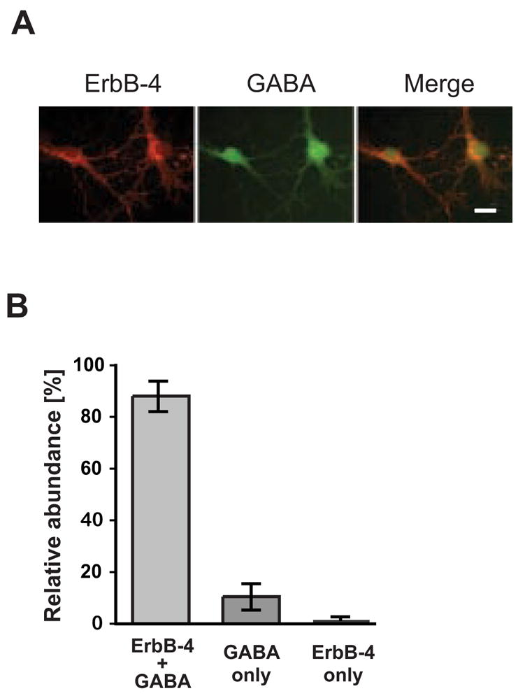
A, Double immunofluorescence labeling of DIV 14 hippocampal neurons with antibodies against ErbB-4 (red) and GABA (green). Scale bar = 20 μm. B, Quantitative analysis of co-expression of ErbB-4 and GABA. The sum of all cells positive for ErbB-4 or GABA was set as 100%. Data represent the mean ±SD of 1004 cells counted in 5 independent experiments.