
Figure 2. Regulation of ErbB-4 and PSD-95 expression in developing hippocampal neurons.
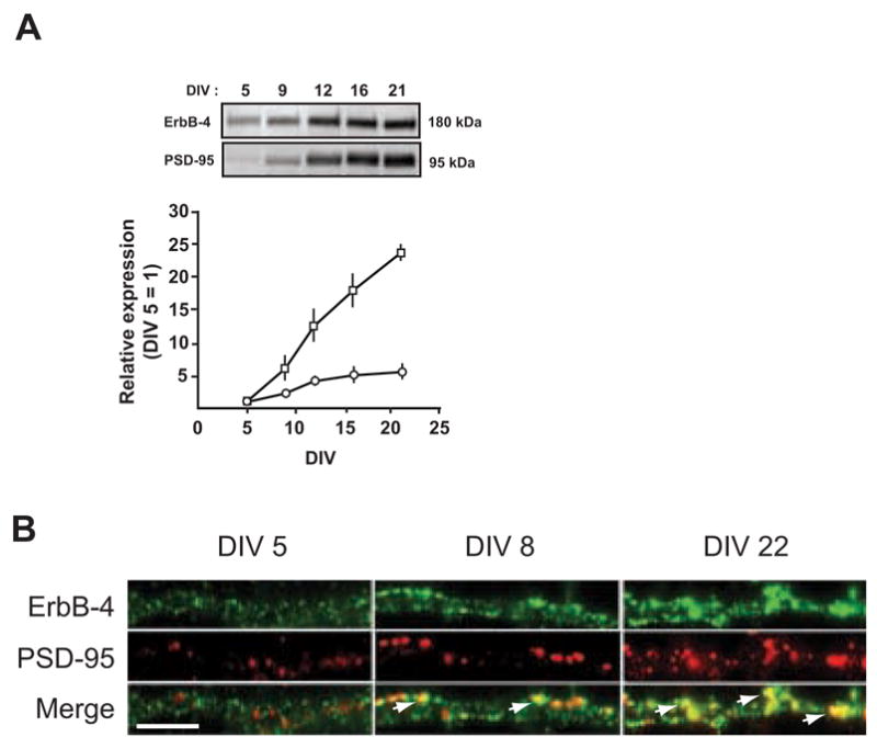
A, (Top) Representative Western blots of whole cell lysates from DIV 5 – 21 neurons, probed with antibodies against ErbB-4 and PSD-95. Equal amounts of protein, as determined by a colorimetric protein assay (Bradford), were loaded in each lane. (Bottom) Quantitative analysis of Western blots. Relative expression levels of ErbB-4 (open circles) and PSD-95 (open rectangles) at DIV 5 were arbitrarily defined as 1. Each data point represents the mean ± SD of three independent experiments. B, Double immunofluorescence of ErbB-4 (green) and PSD-95 (red) in dendritic processes at DIV 5, 8, and 22. Arrows indicate areas of colocalization (yellow) between ErbB-4 and PSD-95. Scale bar = 5 μm.