
Figure 3. Surface expression of ErbB-4 in young and mature hippocampal neurons.
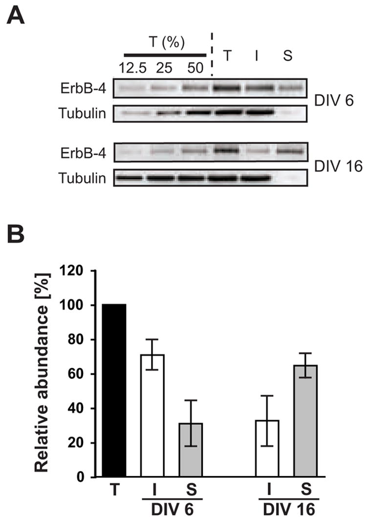
A, Representative Western blots of surface-biotinylated DIV 6 and DIV 16 hippocampal neurons probed with a polyclonal antibody against the carboxyl terminus of ErbB-4. To control for linearity, 12.5%, 25%, and 50% fractions of the total protein input (T) before biotin / streptavidin pulldown were loaded in the first three lanes. I, supernatant after fractionation representing unbiotinylated internal proteins; S, pulldown fraction representing biotinylated surface proteins. To affirm that biotinylation was restricted to surface proteins, blots were re-probed with a monoclonal antibody against a cytosolic marker (tubulin). B, Quantitative analysis of ErbB-4 surface expression. ErbB-4 levels in total input fractions were set as 100%. Bars represent the mean ± S.D. from 7 and 6 independent xperiments for DIV 6 and DIV 16, respectively.