
Figure 7.
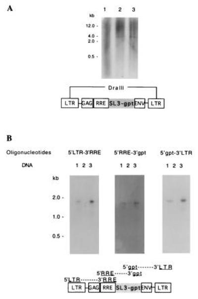
Analysis of the V653gpt vector in the transduced cells. (A) Total DNA from HeLa-T4 cells (negative control, lane 1), transduced HeLa-T4 cells (lane 2), and plasmid V653gpt mixed with HeLa-T4 cellular DNA (positive control, lane 3) were analyzed by Southern blot hybridization after DraIII digestion. DraIII cuts the vector twice, releasing a 4-kb fragment as shown on the diagram. (B) PCRs using the three pairs of oligonucleotides shown on the V653gpt vector diagram were carried out on genomic DNA extracted from transduced HeLa-T4 cells (lanes 1), Ψ422 cells (negative control, lanes 2), or V653gpt plasmid (positive control, lanes 3). The PCR fragments were separated by electrophoresis on a gel, blotted, and hybridized with an HIV-1 LTR probe (oligonucleotides 5′LTR-3′RRE and 5′gpt-3′LTR), or a gpt probe (oligonucleotides 5′RRE-3′gpt).