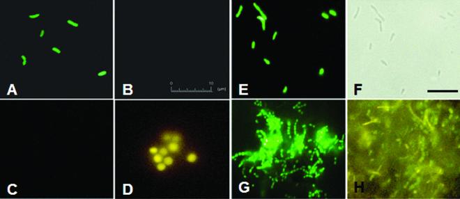
FIG. 3.
Epifluorescence micrographs showing the use of DNA MBs and PNA MB to detect bacterial and archaeal cells. Pure culture of E. coli (A and B) and M. acetivorans (C and D) were fixed by using 4% paraformaldehyde and were incubated with DNA MB Bact0338 (A and C) and DNA MB Arch0915 (B and D). The 10-μm bar in panel B also applies for panels A, C, and D. Fluorescence micrograph of pure culture of E. coli hybridized with PNA MB Bact0338 (E) and corresponding phase-contrast image (F). Fluorescence micrographs of biomass from UASB reactor incubated with PNA MB Bact0338 (G) and DNA Bact0338 and Arch0915 MBs (H). The bar in panel F corresponds to 10 μm and also applies for panels E, G, and H.

