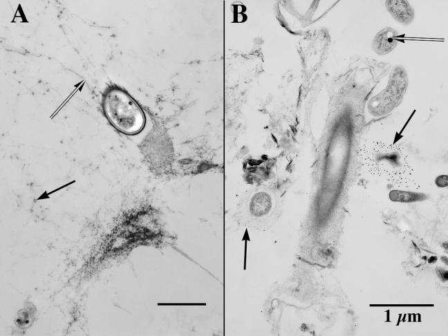
FIG. 3.
(A) TEM image showing a bacterium held within a biofilm by a very porous (water-rich) matrix of fibrillar EPS, fibrils (double arrow) that cross-connect bacteria, and other colloids. Note the electron-opaque nanoscale agglomerations (arrow) on parts of some fibrils. The scale bar on this image represents 500 nm. (B) TEM image of a portion of biofilm that shows several distinctive bacterial morphotypes (including a sheathed filamentous bacterium, several naked gram-negative bacteria with their enclosed storage granules [double arrow], and an irregularly shaped bacterium surrounded by probes that consist of Ulex europaeus lectins attached to colloidal gold spheres [arrow]). Both images were derived by a double-fixation (glutaraldehyde-ruthenium red followed by osmium tetroxide-ruthenium red) protocol, which preceded an embedding in a low-viscosity epoxy resin.

